Nanotechnology in Medicine
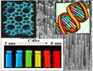
| Many years have passed since 1959 when physicist and Nobel prize winner Richard Feynman proposed the idea of using structures from a nanometer to 100 nanometers size as tiny machines transforming our lives. What is now known as molecular nanotechnology has existed in nature all along. Enzymes are natural molecular nanotype machines, that attach to individual reactant molecules from the surrounding solution and, as a result of precisely orienting themselves with respect to each other in a protected environment, catalyze reactions in this highly specific manner at very high speeds and under body temperatures. If nature can produce the biochemical capabilities of living cells , molecular engineers may be able to accomplish comparable and novel results guided by the examples provided by living systems. Success of nanotechnology in medicine can be broken down to two major areas namely diagnostics and therapeutics. In both areas nanotechnology holds the promise of diagnosis and therapy that is tailor made for each individual. Recently Professor Charles Leiber of Harvard described using arrays of silicon-based nanowire devices to detect certain cancer proteins in the blood at very low concentrations. His group doted the silicon nanowires with monoclonal antibodies specific for the cancer proteins. When the actual protein was present it would attach to the monoclonal antibody and this would cause a charge change in the nanowire and that charge acted as a gate to fire a transistor and record the concentration. This microtransistor allowed for the unique ability to detect very low concentrations.The markers included CEA and PSA proteins which are useful in clinical cancer practice. Similar types of arrays have been developed to find genetic mutations in certain cancer genes while others using tiny gold nanoparticles help identify sequences within DNA. Progress has also been made in direct tumor detection. For example, John Fragioni of Beth Israel in Boston has used quantum dots to image early cancer cells in the lymph nodes of animals.The particles injected in the animal were strong emitters and absorbers of infared light. They quicly would localize, via attached antibodies, in an abnormal lymph node. Then by shining light on the skin external to the node would cause it to light up. For deeper tumors magnetic dots can be used and then scanned with MRI. One problem may be the potential toxicity of various particles and such potential problems can be overcome but must be carefully investigated. The other area is therapeutic methods. Tomorrow's pharmaceuticals could be programmable machines with a range of decision-making and action capabilities. Researchers from Northeasten University are linking chemotherapy with nanoparticles attached to antibodies which can just go to the abnormal cells surface and releasing the drug there and this avoiding the side effects of hitting normal cells. Others have the nanoparticles find the tumor and then use infared light to make these particles cook and destroy only the abnormal tissue. Much remains to be done; however, nanotechnology allows us to get down into the molecular level and to focus in on a diagnostic or medical problem, adjust for individual variations and check for success or error while not being perturbed by or perturbing other cells or structures.11/25/2005 | ||||||||||||||||||||
see comments or
post your comment
see previous blog
see next blog